Original Articles: 2023 Vol: 15 Issue: 2
In-vivo Amelioration and Neuroprotective Effect of Morus alba Fruit Extract on Formaldehyde Induced Neurotoxicity as an Alzheimers Model in Zebrafish Brain
Kavitha G Singh*, Aparna R Nair, Hamsa RK
Department of Biochemistry, Mount Carmel College, Bangalore, India
Received: 11-Oct-2022, Manuscript No. JOCPR-22-76885; Editor assigned: 13-Oct-2022, PreQC No. JOCPR-22-76885 (PQ); Reviewed: 27-Oct-2022, QC No. JOCPR-22-76885; Revised: 09-Jan-2023, Manuscript No. JOCPR-22-76885 (R); Published: 16-Jan-2023
Abstract
The fruit of Morus alba, also known as the mulberry, is a rich source of phytochemicals like anthocyanin, quercetin, cholinergic acid, and resveratrol, of which the aglycone found in anthocyanin and cyanidin-3-o-d-glucopyranoside (c3g) has been identified to demonstrate free radical scavenging, inflammatory suppressing and neuroprotective action. The fruit extract has a range of biological actions, with the neuroprotective, antioxidant and anti-inflammatory effects being particularly beneficial in situations like neurodegeneration. Alzheimer’s disease and other NDDs are characterized by the build-up of ROS species, which directly speeds up the development of tau proteins and neural plaques which result in loss of memory and other cognitive abilities. One of the key contributors to the development of Alzheimer's disease has been identified as age and the accumulation of harmful substances in the body over time. Formaldehyde (FA) metabolizes in the body and stabilizes the formation of tau proteins, resulting in a vicious cycle. Hence, there is a need to develop a medication that can diminish or completely prevent the development of AD rather than only reducing its symptoms. In our study, we look at the curative benefits of Mulberry fruit extract on Zebrafishes by analyzing their behaviour in different courses of experimentation and evaluating histopathologically sections.
Keywords
Morus alba; Anthocyanin; Alzheimer’s; Formaldehyde; Neurodegenerative diseases
Introduction
Alzheimer’s disease is a brain ailment that causes memory and thinking skills to deteriorate over time, eventually leading to death. A reduction of thinking ability, vision issues, impaired reasoning and mild cognitive impairment as the disease progresses. According to a poll conducted in the United States, Alzheimer’s disease is currently the sixth largest cause of death among the elderly. As of 2020, there are over 6 million Americans aged 65. As a result, this figure is expected to nearly triple to 14 million by 2060 [1].
Greater memory loss and other cognitive challenges, such as wandering and getting lost, questioning repeatedly, prolonged time to complete typical everyday chores, etc. are experienced as Alzheimer’s disease advances. As disorientation and memory loss worsens, it gets harder for people to identify their loved ones. They could have trouble learning new things, delusions, paranoia, hallucinations and impulsive behaviour. In their most critical stages, someone will need to take care of them.
The most well-known risk factor for Alzheimer’s disease is advanced age. Scientists have made enormous strides toward understanding Alzheimer's disease in recent years and the impetus is still building. Even still, researchers are still unsure of the exact causation of Alzheimer's in most cases. Adding on to the factors, diet, lifestyle and numerous environmental factors all have a role in the cause of Alzheimer’s illness progression.
Industrialization contributes to an elevation in environmental contamination, with industrial air, commercial pollutants, and combustion of organic chemicals posing numerous health risks. Exposure to organic chemicals causes acute and chronic toxicity, imbalance of various metabolisms and physiological functions in the body leading to degenerative diseases [2,3].
In the present day lifestyle, exposure to organic chemicals used in resins, plastics, detergents, dyes, natural gas and medicines, causes amyloid protein aggregation on neuronal cells, suggesting that chemical exposure may cause AD-like diseases [4].
According to a study, formaldehyde is formed due to forest fires, industrial emissions, incineration and fuel combustion. It is also present in building materials, press fabrics, paints, body and hair care products. People working in industries, laboratories, health care professionals and automobiles are exposed to absorbing formaldehyde containing substances via the skin or breathing formaldehyde gas or vapor from the air.
When metabolized in the body, the aggregation of beta amyloid peptides occurs, causing cognitive impairment because of tau protein phosphorylation which eventually leads to dysfunction and even apoptosis [5-7]. Even a low concentration of 0.01% formaldehyde can damage the senile plaques, intracellular ROS accumulation in the body, when exposed repeatedly, develops lack of focus, memory loss and sleeplessness. Long term exposure to formaldehyde at airborne levels can impair the development of seizures and loss of cognitive function, showing reduced balance, vibration sense, color discrimination, and blink reflex latency [8].
Although there is currently no cure, tremendous progress has been made in recent years in discovering treatments to delay the onset of the disease. According to several scientific studies conducted on various levels such as cells, animals and people, consuming berry fruits have positive effects on the brain and may help prevent age related memory loss and other changes. Due to the high levels of antioxidants found in them, which can alter neuron signaling to protect cells from damage induced by dangerous free radicals and enhance both motor coordination and cognition, inflammation in the brain can also be prevented. In our study, we focused on the genus Morus and species of Morus, which is alba of the berry family to investigate the neuroprotective impact observed.
Mulberry (Morus alba) contains numerous bioactive that provide health benefits that go beyond basic nutrition and have a diverse group of phytochemicals by modulating signaling pathways involved in inflammation, cell survival, neurotransmission and neuroplasticity, berry fruits can help prevent age related neurodegenerative diseases and improve motor and cognitive functions [9]. They are of great biological and pharmacological interest and play a crucial function in inflammatory cytokine signaling used to cure dizziness, sleeplessness and premature aging as an ancient medicine [10]. In our study, we determine the extent of mulberry in counter effect to Alzheimer’s disease using the Zebrafish as a model.
As there is a need to follow ethical considerations for conducting and reporting animals for research purposes, we utilize Zebrafish as a replacement for human studies, they share common physiological features’ and genomically related genetic disorders by sharing homology with most human sequences, which paves way for the study of neurodegenerative diseases.
Zebrafish (Danio rerio) is a tropical freshwater fish, an excellent model for human disease research as they share common physiological features by sharing 50%-80% homology with most human sequences and genomically linked genetic problems, paving the way for translational neurodegeneration research. Having a small life span in development with a high reproductive rate like humans, it demonstrates several higher order activities like memory, conditioned reflexes, and social behaviour. Zebrafish brain organization is like that of humans, sharing structural properties, as well as neurotransmitter circuits. Given with ease in pharmacological manipulations, its complex neurological system, and wide behavioral range, it is an excellent transitional model to study the effects of psychotropic compounds [11]. Additionally, it is an optimistic model for visualizing, experimentally manipulating, and interpreting the molecular basis of human neurodegenerative diseases in recent times [12].
By examining the histopathological and behavioral alterations in the Zebrafish brain, the current study sought to evaluate the formaldehyde induced neurotoxicity in Zebrafish and the neuroprotective effect displayed by Morus alba fruit extracts. The similarity between the neurotransmitter systems of Zebrafish and those of humans makes this model a good method for studying neurotoxicity. It is also inexpensive, time saving and ethical.
As the adage goes, prevention is better than cure. By including naturally occurring sources in our diets, we can significantly slow the progression of the major diseases that are prevalent due to a variety of factors in today’s society’s lifestyle. This is safer for the body than consuming conventional synthetic medications.
MATERIALS AND METHODS
Chemicals
All the chemicals were of the highest grade. Methanol, 7,11b-dihydro-6H-indeno [2,1-c] chromene-3,4,6a,9,10-pento,disodium;2-(2,4,5,7-tetrabromo-3-oxido-6-oxoxanthen-9-yl) benzoate, distilled water, ethanol.
Experimental animal model
The experimental animal model used was Zebrafish (Danio rerio), which were obtained from a local fish dealer in Bangalore. The fish used in the study were 2 months-3 months old and of the same size. Fishes were maintained in a 500 ml water tank for acclimatization to the laboratory conditions for a week and supplied with external oxygen. Commercially available fish feed was used to feed the fish once a day, as part of their regular diet. The temperature of about 24°C-26°C, the humidity of 55%-60%, the photoperiod maintaining a 12:12 hour light dark cycle and the pH of 7.0-8.0 were all maintained in a controlled setting.
Experimental procedure
The experimental test was set up for a month and all histological examinations and behavioral analyses were performed regularly throughout that time. As an experimental model, 30 fish were chosen and separated into two groups: control (i)and diseased (ii). Control fishes were maintained in a normal environment supplemented with a normal diet.Diseased fishes were exposed to fresh 10% formaldehyde solution prepared in distilled water once a day for a knownperiod. During the exposure period, all behavioral parameters were evaluated.
Following the completion of the exposure, the diseased fish were sacrificed to determine the disease’s course. The treatment was administered based on the results of the disease’s prevalence through histological examination. The fishes were treated with Morus alba extract separately in distilled water at a concentration of about 0.5 ml/500 ml for the next 10 days. The fish were tested after 10 days of treatment for further behavioral analysis.
Brain tissue for histological examination
By generating hypothermic shock on cooled ice, the fishes were sacrificed. The fish’s cranial region was severed, separating it from the body. To obtain the brain tissue, the skin, eyes, mouth and meat of the head were carefully peeled away.
Histological study
Haematoxylin and Eosin (H and E) staining method: The histochemical tissue pathology of cationic and anionic components in tissue sections was visualized using the haematoxylin and Eosin (H and E) staining technique.
The slides containing the tissue sections were placed in a metal staining rack. The slides were treated with haematoxylin for 10 seconds followed by washing. Again, the slides were immersed in eosin stain for 30 seconds and washed to make the slides clear. The slides were dehydrated in a series of progressively stronger alcohol solutions of 50%, 70%, 80%, 95% and 100% respectively in a staining jar. The sections were visualized under the compound microscope.
Behavioural study
Behavioural experimental setup: The goal of this study was to evaluate behavioural alterations in swimming patterns caused by formaldehyde exposure. A 500 ml tank containing 10% formaldehyde solution was taken by the fish.
To notice the effect of exposure on fishes and the behavioural phenotypes in the fishes during the exposure period, we observed the fishes with various parameters in periodic intervals every 2 hours after the exposure for a known time. We recorded and interpreted the behaviour of the fishes concerning the exposure period by dividing it into (i) Onset of exposure, (ii) The middle of exposure, (iii) The culmination of the exposure and (iv) Post mulberry treatment.
RESULTS
Histopathological analysis
Brain cells have a distinct shape and structure; however, during Alzheimer’s disease, a distinct alteration occurs, which can be observed using several staining techniques.
H and E staining
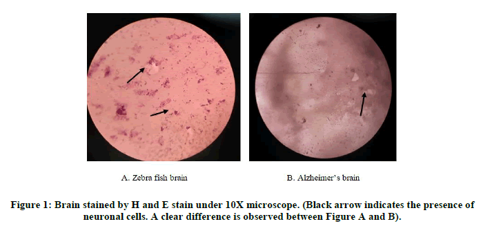
A basic morphological stain that has been used to differentiate between the nucleus and cytoplasmic contents of neuronal cells. In H and E nuclei and other basophilic structures are stained blue whereas cytoplasmic contents and acidophilic structures are light to dark red.
On observation, a reduced number of neuronal cells were observed in Alzheimer’s fishes in comparison to control fishes indicating characteristic destruction of cells. Additionally, cells present within the diseased appeared distorted and shapeless (Figure 1).
Behavioural analysis: We observed the behavioural aspects of the fishes as follows.
During the onset of exposure
Alarm reaction: The fish showed increased velocity, inconsistent locomotion and fast direction changes to escape from the exposure.
Aggression: The fishes showed behaviors like fin elevation, undulation, biting and chasing the other fishes as a defense mechanism which indicates their aggression.
Bending and rotation: The fishes swimming in a laterally bent position, sometimes in a circular direction along with twitch and spasms movements with a muzzle velocity.
Jittery swimming and seizures: The fish is attributed to repeated strained and anxious motions with poor swimming abnormalities which is a sign of jittery movements that eventually results in reflexive shivering leading to seizures.
In the middle of the exposure
Akinesia and ataxia: The fishes showed incoordination of bodily movements with hypo locomotion with a drooly tail associated with motor incoordination showing a stated body posture.
Buoyancy dysregulation: The fishes showed a disability to remain at a constant elevation of water levels, which forces physical effort in inclined swimming and tilting.
Coasting and corkscrew swimming: The fishes showed inactive sliding without bodily movements by the usage of pectoral fin alone in decreased swim rate in a different direction indicating submissive behaviour.
At the culmination of exposure
Surfacing or floating: The fish showed passive swimming either by dwelling at the surface of the water or drifting in the centre of the tank indicating sedation.
Lethargy: The fishes showed behaviour as an indication of chronic distress and illness by behaviours such as decreased locomotor activity, reduced escape response and staying close to the bottom of the tank.
Sickness and lethargy: The inactiveness which inhibits the feeding hypo activity, exhibits misery and suffering and lethargy is sedentary to the bottom of the tank.
Paralysis: Complete cessation of all movement, including the eyes, gills/operculum and fins, followed by motor dysfunction and an aberrant posture and vertical float sometimes leading to death.
Post mulberry treatment
Appetitive olfactory behaviour: Increased foraging behaviour, attractiveness and nibbling were observed in relation to food that had been suppressed owing to illness by modifying the appetite.
Counter swimming approach: In contrast to the patterns observed throughout the exposure phase, persistent swimming movements without changes were displayed for an extended period.
Withdrawal related behaviour: A specific behaviour shown because of the discontinuance of target chemical induction, generally marked by enhanced anxiety/fear like behaviour.
Congenial light exposure: Reportedly, fishes showed more phototaxis response post-treatment, which preferred to be photo negative during the induction.
Complex behaviours: Tremor behaviour associated with seizures, startled behaviour, trance like swimming patterns, and negative thigmotaxis all decreased in proportion to the behaviour displayed by the fishes during the exposure.
DISCUSSION
Altering behaviour and diminishing memory are the major manifestations of AD. To evaluate and understand this process of culmination in variation differing parameters were analysed during the entire course of exposure. Initially, when the fishes were acclimated to FA they displayed extreme alarm response, inconsistent swimming patterns, rotational swimming, biting and chasing other fishes, all of which were clear indications of aggression. In the middle of exposure, the fishes were seen to have buoyancy dysregulation with irregular body movement displaying droopy tail, slide motion of fishes and reduced swim rate this portrays the submissive nature and prognosis of the disease, affecting the ability to think and direct different organs to function. These parameters clearly indicated the presence of AD and to conclusively prove its presence histopathological analysis was performed on the fishes by H and E stain which indicated the destruction of neuronal cells in the diseased brain. Before traversing the fishes into the culmination of exposure a group of fishes was taken and treated with mulberry, it was observed that fishes that were at the middle of exposure when withdrawn from treating with FA and treated with mulberry displayed significant improvement in behaviour, displaying increased thigmotaxis, foraging patterns, phototaxis but at the same time showed a minor level of the trigger and trance like behaviour. The other group which was being continued to be exposed by FA showed increasing surfacing and floating patterns, they showed little to no movement with prominent drowsiness.
CONCLUSION
In conclusion, we were able to identify that increased exposure to formaldehyde resulted in altered behaviour and cognitive abilities which was directly linked to Alzheimer’s. By analysing the behavioural parameters, we were able to demarcate the stages of exposure and the altered behaviour displayed during different points.
Three major points of exposure were determined by altering patterns; in the second stage of exposure AD like changes were increasingly identified and were followed up with histopathological analysis which showed a positive result. To determine the neuroprotective potential of Morus alba a group of fishes was divided amongst them and treated with the extract. After analysis, it could be understood that mulberry fruit administration could reduce behavioral abnormalities associated with AD and therefore could offer a potential therapy.
REFERENCES
- Matthews KA. Alzheimers Dement. 2019;15(1):17-24.
[Crossref] [Google Scholar] [PubMed]
- Alissa EM, Ferns GA. J Toxicol. 2011;2011:1-21.
[Crossref] [Google Scholar] [PubMed]
- Tchounwou PB, Yedjou CG, Patlolla AK, et al. Exp Suppl. 2021;101:133-164.
[Crossref] [Google Scholar] [PubMed]
- Kawahara M, Kato-Negishi M. Int J Alzheimers Dis. 2011;2011:1-17.
[Crossref][Googlescholar] [PubMed]
- Li F, Gong S, Zhang H, et al. Environ Res. 2020;184:109318.
[Crossref] [Google Scholar] [PubMed]
- Li ZH, He XP, Li H, et al. Zool Res. 2020;41(4):444-448.
[Crossref] [Google Scholar] [PubMed]
- He R, Lu J, Miao J. Sci China Life Sci. 2010;53(12):1399-1404.
[Crossref] [Google Scholar] [PubMed]
- Kilburn KH. Arch Environ Health. 1994;49(1):37-44.
[Crossref] [Google Scholar] [PubMed]
- Subash S, Essa MM, Al-Adawi S, et al. Neural Regen Res. 2014;9(16):1557-1566.
[Crossref] [Google Scholar] [PubMed]
- de Oliveira AM, do Nascimento MF, Ferreira MR, et al. J Ethnopharmacol. 2016;194:162-168.
[Crossref] [Google Scholar] [PubMed]
- Michael Stewart A, V Kalueff A. Curr Neuropharmacol. 2012;10(3):263-271.
[Crossref] [Google Scholar] [PubMed]
- Xi Y, Noble S, Ekker M. Current Neurol Neurosci Rep. 2011;11(3):274-282.
[Crossref] [Google Scholar] [PubMed]

